
Compressive Sampling OCT
Acquiring three dimensional image volumes with techniques such as Optical Coherence Tomography (OCT) relies on reconstructing the tissue layers based on reflection of light from tissue interfaces. One B-mode scan in an image is acquired by scanning and concatenating several A-mode scans, and several contiguous slices are acquired to assemble a full 3D image volume. In this work, we demonstrate how Compressive Sampling (CS) can be used to accurately reconstruct 3D OCT images with minimal quality degradation from a subset of the original image. The full 3D image is reconstructed from sparsely sampled data by exploiting the sparsity of the image in a carefully chosen transform domain. We use several sub-sampling schemes, recover the full 3D image using CS, and show that there is negligible effect on clinically relevant morphometric measurements of the optic nerve head in the recovered image. The potential outcome of this work is a significant reduction in OCT image acquisition time, with possible extensions to speeding up acquisition in other imaging modalities such as ultrasound and MRI.
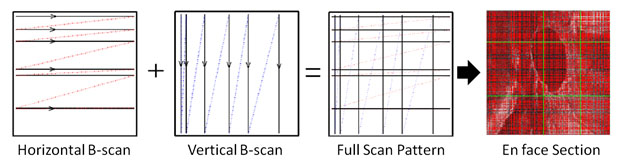
Human Optic Nerve Head (ONH) volume from a 830nm Spectrometer-based OCT system was used as model data to evaluate CS recovery technique. The sparsely sampled OCT image volume that would be acquired by our proposed method of skipping horizontal and vertical raster-scan lines (Figure 1) was simulated by applying a binary restriction mask to the data.
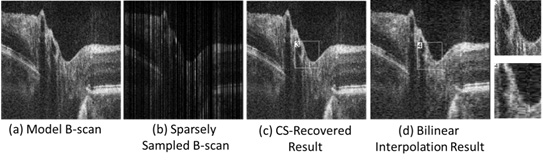
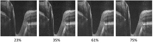
Representative results from the CS OCT are presented in Figure 2 and Figure 3. Figure 2 compares CS recovery technique to bilinear interpolation on a volume with 53% missing data, and Figure 3 presents representative B-scans recovered with CS in volumes with 23, 35, 61 and 75% missing data.
Fewer total B-scans are required for CS-based recovery of the OCT volume, reducing the acquisition time with minimal degradation in image quality. The reduction in the OCT scan time limits the discomfort of a subject during the scan and reduces the amount of motion artifact. Image reconstruction with CS preserved the smoothness of sharp features in the optic cup, whereas standard bilinear interpolation resulted in staircase effect and loss of detail. The Compressed Sensing scan pattern is compatible with existing OCT acquisition hardware, and has the potential to reduce the acquisition time in applications such as retinal imaging where the Ascan rate is limited by the maximum permitted optical exposure.
Reference
E. Lebed, P.J. Mackenzie, M. V. Sarunic, M.F. Beg, "Rapid Volumetric OCT Image Acquisition Using Compressive Sampling", Opt. Express 18 (20), 21003-21012 (2010).