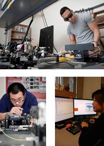
1) Biomedical Optical Imaging - Dr. Marinko Sarunic
Background: Medical imaging provides the ability to look inside biological samples non-invasively. Techniques like CT, MRI, and ultrasound are designed for whole body imaging, and microscopes are used for looking at cells. There is a missing layer between them for studying small biological systems which are on the order of millimeters. A newly emerging technique for tissue imaging studies is called Optical Coherence Tomography (OCT) which is similar in principle to ultrasound, but uses light instead of sound waves. OCT systems have resolution and imaging depth intermediate between microscopy and high frequency ultrasound, filling an important technological gap. OCT is widely used to image the retina in human, and also as a tool to augment studies in developmental biology.
Objectives: The purpose of this project is to learn about optical medical imaging using Optical Coherence Tomography. Through directed study, the Undergraduate Research Assistant will learn basic concepts in optics and use them to build a primitive OCT system in order to gain understanding of operation. Next, the Undergraduate Research Assistant will use a real OCT system for image acquisition on biological systems. Basic medical image processing with MatLab will be used to improve the image, and 3D visualization software will be used for presentation. The work on the project is diverse, ranging from optics, to programming, image processing, and 3D display. Concentration in one aspect of the project will be determined by the Undergraduate Research Assistant’s strengths and interests.
Skills: Familiarity with MatLab and C++ is preferred.
2) Three dimensional OCT image processing and visualization using GPU -- Dr. Marinko Sarunic
Background: Biomedical optical imaging is a rapidly emerging field for medical diagnostic imaging. Optical Coherence Tomography (OCT) is a method of obtaining high resolution sub-surface cross sectional images in biological samples. Modern OCT systems have heavy computational requirements in order to reconstruct images from unprocessed data. New OCT systems permit rapid acquisition of raw data faster than can be processed with a standard desktop computer, and high speed hardware solutions are required for real time image display.
Objectives: This research project will have a student investigate the initial design phases for creating a GPU accelerated implementation of real time OCT volume acquisition, leveraging both the increased parallelism and pipelining possible in hardware (spatial computing). The initial modules the student will be designing will focus on furthering the existing work on pre-processing and frequency domain transforms (Fast Fourier Transforms) for real time visualization of data. Additional work may include volume rendering.
This project will strengthen the student’s software design skills via the implementation of a medical imaging application. Understanding optics is not required, but completion of the Advanced Digital System Design course would be a bonus.
Skills:
- high-level language programming skills (eg C, C++, C#) are a must
- Completion of ENSC 381 (signals and systems) will facilitate the student’s understanding of the processing algorithms
- Experience with CUDA would be an asset

